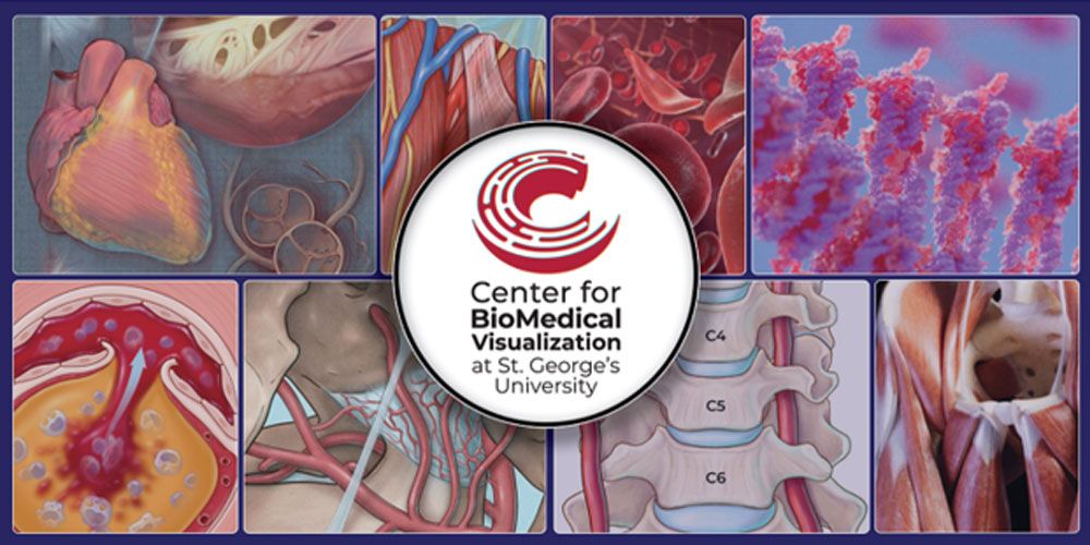
Medical illustration plays an important role in medical education. From the first anatomy lecture to the final clinical rotation, students are asked to understand structures, systems, pathways, and disease processes that are often difficult to picture from words alone. At St. George’s University School of Medicine, our team of faculty medical illustrators in the Center for BioMedical Visualization help bridge that gap by creating custom educational visuals designed specifically for the way students learn.
Our medical visualization work supports lectures, research projects, posters, interactive teaching materials, and student learning resources across the School of Medicine curriculum. Whether we are building a 3D model of an anatomical structure, designing a selective-disclosure PowerPoint animation, creating a research illustration, or scanning cadaveric material for teaching, the goal is always the same: to simplify complex medical information and make it easier to retain.
The idea for the Center began in 2012 with Dr. Marios Loukas, Dean of the School of Medicine (SOM) and University President, who proposed bringing medical illustrators to SGU to support SOM’s Department of Anatomical Sciences’ growing research output with detailed illustrations.
“The goal for the Center for BioMedical Visualization was to not only deliver to students high-quality learning materials, but to embrace new technologies and teaching tools on campus and throughout the wider SGU community.”
—University President Marios Loukas, MD
Why medical illustration matters in medical education
Medical students are expected to learn an enormous amount of information in a short period of time. Anatomy education, embryology, histology, pathology, physiology, and other clinical concepts all require students to think spatially and make connections between form and function. Traditional lecture slides and textbook images are helpful, but custom imagery that links directly to SGU’s established learning objectives and curriculum content is where our work is most effective. A well-designed image can simplify a difficult topic, emphasize what matters most, remove distractions, and guide the learner through a concept, step by step. At SGU, our team creates visuals that are built around the curriculum, the instructor’s goals, and the needs of students. They are educational materials created with a clear purpose rather than just a visual accessory. In many cases, custom visuals can also help shorten lecture time because an idea that might take several minutes to explain verbally can sometimes be understood more quickly through a clear diagram, animation, or interactive model.
How medical illustration supports student learning at SGU
Our team works closely with faculty to develop custom imagery for student lectures, educational modules, research posters, publications, and interactive teaching materials.
With specialties ranging from research publication illustrations to 3D medical animation and modeling, the team works on a wide array of projects, including:
- 2D and 3D animations explaining biological processes,
- Interactive learning modules and virtual anatomy tools,
- Posters, presentations, and infographics for conferences, especially for faculty,
- Custom illustrations for faculty research and peer-reviewed publications,
- Educational outreach materials for community health initiatives, and
- Dissection and instruction in both undergraduate and graduate anatomy labs.
Each modality serves a different educational purpose for our students. A 2D illustration may be the best way to show a clean, simplified overview of a nerve pathway. A 3D model may be better for helping students understand spatial relationships in anatomy or protein mechanisms within a cell. A well-timed and clear animation can reveal information gradually, allowing students to build understanding one layer at a time rather than getting overwhelmed by a fully labeled diagram displayed all at once.
How interactive 3D models support anatomy education
Interactive 3D models are especially valuable in anatomy education because they allow students to rotate, inspect, and study structures from multiple angles. This is important because the human body is not flat. Students need to understand how structures relate to each other in space, and 3D visualization gives them a way to explore those relationships more effectively.
For example, first-year SGU medical students in the anatomy lab use custom interactive 3D models directly at the wet lab stations. While observing real dissected specimens, they use the models like a digital map, deepening their understanding in real time.
How medical illustration supports medical research and communication
Medical illustration also plays an important role in student and faculty research. Research posters, journal figures, and presentation graphics should communicate ideas quickly and accurately and will often garner more attention to a given article. Our team helps students and faculty transform complex findings into visuals that are clear, professional, and scientifically grounded.
For students, this support can make a meaningful difference. A strong research poster or figure can help them explain their work more confidently at conferences and better communicate with scientific and clinical audiences. These are skills that extend beyond medical school. Physicians must constantly explain complex information to patients, colleagues, and communities. Learning how to explain concepts visually is part of learning how to communicate medicine.
The work of our medical illustration team reflects SGU’s commitment to modern, student-centered education. We use scientifically informed visual communication methods to support how students learn today.
Medical illustration helps students see the bigger picture
Medical illustration is about storytelling. It helps students navigate and organize complex information and connect what they are learning in the classroom to the practice of medicine. This type of educational support can make challenging material more approachable and meaningful. At SGU School of Medicine, our faculty medical illustrators are part of that support system. We help students engage more deeply with their education and build a stronger foundation for their future careers.
See how you’ll learn at SGU
Medical illustration is just one of the ways SGU supports students throughout their medical education. Explore the 4-Year MD Program to learn more about the curriculum, learning environment, and path from Basic Sciences through clinical training.