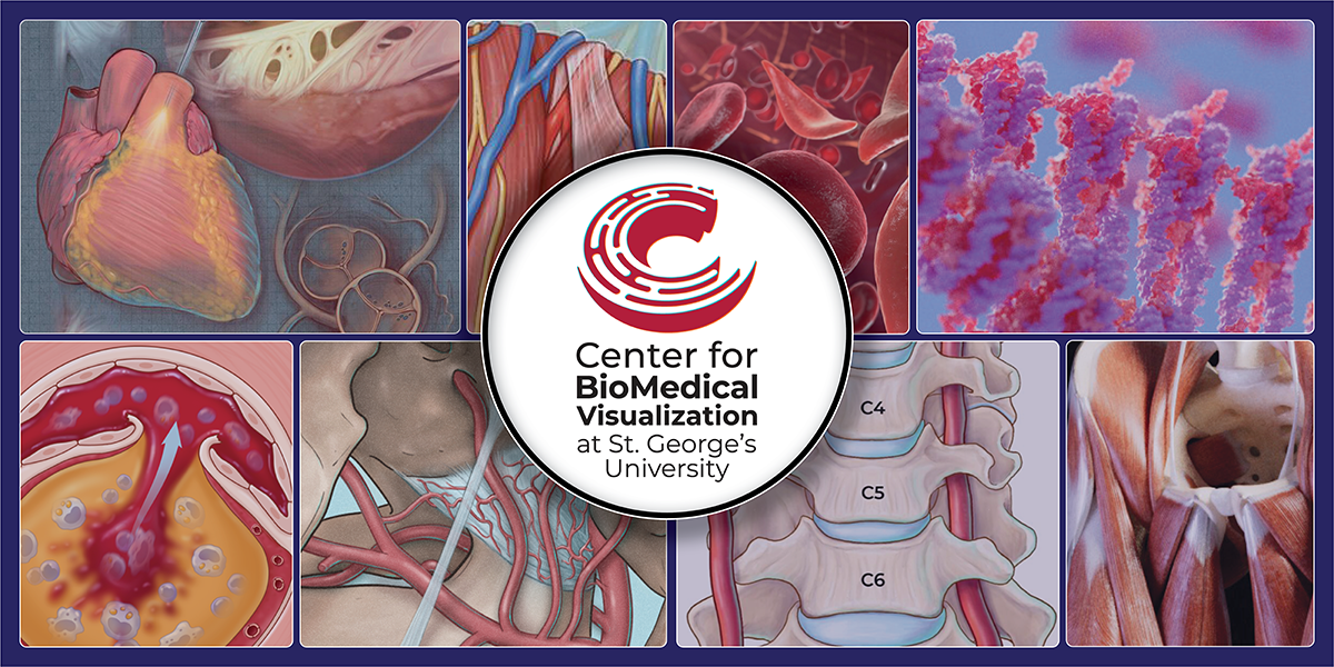
Tucked behind the scenes of lecture halls and lab manuals, a team of talented faculty at St. George’s University (SGU) is bringing science to life—one brushstroke at a time. The Center for BioMedical Visualization is where creativity meets clinical precision as medical illustrators transform complex anatomical concepts into vivid, digestible visuals that jump off the page and into students’ minds.
From intricately rendered 3D models to dynamic animations, these visual storytellers are not just adding color to the curriculum—they’re shaping the way future doctors learn, understand, and retain critical knowledge.
“The Center elevates the learning experience of the students through exciting visual content. In collaboration with other faculty, we create learning materials that condense large amounts of complex information into clear, easy-to-understand illustrations, animations, and models,” explained Sue Simon, MS, CMI. Ms. Simon serves as director of the Center and an instructor of anatomical sciences. She is a board-certified medical illustrator with over 20 years of experience and will be beginning her Master of Education (M.Ed.) degree at SGU this fall.
“We meticulously tailor everything we do to be in sync with what our med students are learning. From the start of Term 1 to the conclusion of Term 5, our work is found across the entire student experience,” she continued.
For aspiring doctors, the Center for BioMedical Visualization creates more than just learning aids. As medicine and technology continue to evolve, SGU students can gain an edge by learning through tools that make the curriculum interactive, intuitive, and memorable. It is a resource that builds the clinical confidence needed for real-world success.
It started with an idea
The idea for the Center began in 2012 with Dr. Marios Loukas, dean of the School of Medicine (SOM) and University president, who proposed bringing medical illustrators to SGU to support SOM’s Department of Anatomical Sciences’ growing research output with detailed illustrations.
“The goal for Center for BioMedical Visualization was to not only deliver students with high-quality learning materials, but to embrace new technologies and teaching tools on campus and throughout the wider SGU community.” —University President Marios Loukas, MD
As SGU’s faculty output and student body expanded, so did the need for visual support. By 2015, the Center had grown to a four-person team. Just three years later, that number more than doubled.
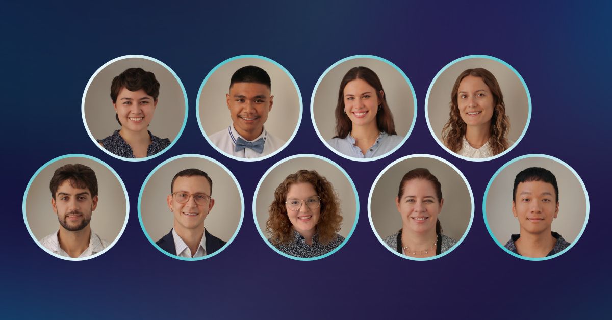
Today, the Center is home to nine illustrators and animators, who are also members of faculty in the anatomy department. The team includes multiple board-certified medical illustrators, all bringing a unique blend of creativity and scientific training to the table. The team includes:
- Sue Simon, MS, CMI, director
- David Nahabedian, MSMI, CMI, assistant director
- Sarah Gluschitz, MA, CMI
- Linden Pederson, MSMI, CMI
- Courtney Brendal, MA
- Janine Murta, MSc
- Max Dragan, BFA
- Sunghwan Bae, MS, MFA
- Jan Bertulfo, BFA
“We strive to always improve our knowledge and skills in order to stay at the forefront of our industry so we can provide the best possible outcomes for our work,” Ms. Simon shared.
With specialties ranging from research publication illustrations to 3D animation and modeling, the team works on a wide array of projects, including:
- 2D and 3D animations explaining biological processes,
- Interactive learning modules and virtual anatomy tools,
- Posters, presentations, and infographics for conferences, especially for faculty,
- Custom illustrations for faculty research and peer-reviewed publications,
- Educational outreach materials for community health initiatives, and
- Dissection and instruction in both undergraduate and graduate anatomy labs.
Pulling the curtain back
What goes into the Center’s illustration magic?
For the team, reliable reference materials are essential for accurate, detailed 3D products, which are gathered through various methods. The team uses strategies like photogrammetry (taking many photos of an object to turn it into a digital 3D model) and extracting models from MRIs and CTs.
“Having the anatomy lab at SGU means we are able to examine structures on those who have given the gift of body donation,” Ms. Simon said.
Then the team runs multiple reviews for quality control including assessing anatomy, ensuring all animated elements interact properly, consulting proper reference materials, and, most importantly, working directly with the project requestors and other SGU faculty for their expertise.
Of all the visual products the team can produce, 3D animations can be the most challenging to get just right as it requires meticulous planning, highly technical software, and a knowledge of physics, anatomy, and motion principles for movements to look believable.
“During the animation phase each scene must be crafted carefully,” said Mr. Nahabedian, assistant director of the Center and anatomy instructor. Mr. Nahabedian, a certified medical illustrator, has been with the Center for eight years.
“Animators must understand 3D modeling, rigging, texturing, lighting, simulation, and rendering—all within the same project, which can be quite the task. Of course, they must also understand the science behind what they are demonstrating. As such, even short scenes can take days or weeks!” he explained.
A 3D animation showing the eukaryotic DNA packaging process. © 2025 Linden Pederson, MSMI, CMI, St. George’s University.
The value in visuals
From the very first term through advanced clinical modules, the Center’s work is embedded across the entire student experience. Whether it’s a detailed animation of a biological process or an interactive 3D model in the anatomy lab, these visuals make dense material more accessible and engaging. It’s not just about making things look good—it’s about making things comprehensible.
Anatomy is one of the clearest examples of this in action.
Understanding the body in three dimensions is essential for future surgeons, radiologists, and general practitioners. However, flat images can only go so far. That’s where the Center’s 3D models come in.
These 3D tools allow students to rotate, zoom, dissect, and explore anatomical structures from all angles, helping them build true spatial awareness. Visual and hands-on learners especially benefit from this approach, as it mirrors how they’ll engage with the human body or animals in clinical settings.
For example, first-year SGU students in the anatomy lab use custom interactive 3D models directly at the wet lab stations. While observing real dissected specimens, they use the models like a digital map, deepening their understanding in real time.
“By providing immersive, highly-detailed visuals, the Center strengthens the connection between theory and practice. Students don’t just memorize. They understand. They see how systems work together. They gain the kind of clinical perspective that typically takes years to develop, and that leads to better-prepared, more capable graduates,” said Dr. Loukas.
For the team, the results are clear: students are more focused, more curious, and more confident in what they are learning with these tools. And that, in turn, helps yield more focused, curious, and confident doctors.
All images © St. George’s University, 2025