Working in pathology is a great path for physicians who are particularly interested in uncovering answers to difficult medical questions. Pathologists are often considered a “doctor’s doctor,” as they’re called upon by their colleagues for diagnostic support in a range of different scenarios.
Assisting in the diagnosis, treatment, and study of diseases, these highly trained professionals will typically specialize in one of the many different types of pathology. One critical subspecialty in the field is cytopathology—a branch of anatomic pathology that involves the study of cells via microscopy.
The diagnostic advancements made possible by cytopathology have paved the way for life-saving treatment plans for a range of different diseases and conditions. Keep reading to examine the details of this important pathology subspecialty.
What is cytopathology?
In order to understand what cytopathology is, you must first know what cytology is. Cytology itself is the study of normal cells—but it is used in conjunction with cytopathology, which is the study of diseased cells. Together, cytology and cytopathology comprise a common diagnostic method used to screen and triage for cancers and other conditions.
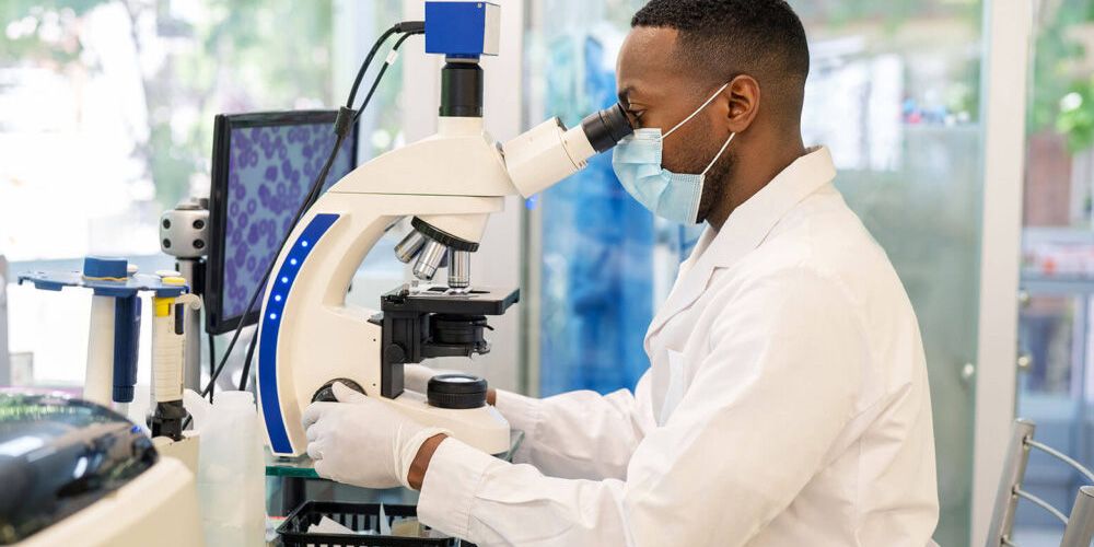
Tests typically examine cells from small amounts of body tissue or fluid under a microscope. Cytopathologists search for characteristics or abnormalities that may indicate a particular diagnosis and/or direct the patient for more specific testing.
There are two main branches of cytology:
Exfoliative cytology is when the cells being examined are either shed by a patient’s body naturally or have been manually scraped or brushed from the surface of their tissue. Examples of samples collected through exfoliative cytology include the following:
- Gynecological samples, such as those gathered from a pap smear
- Gastrointestinal tract samples, such as those collected during an endoscopy procedure
- Skin or mucus samples, such as those scraped from the inside of the nose or mouth
- Respiratory samples, such as those collected from spit and mucus
- Urinary samples, collected from urine
- Discharge or secretion samples, such as those collected from the eye
Intervention cytology is when a specialist must intervene with a patient’s body to collect a sample of cells to test. This is most commonly performed through fine-needle aspiration, which is when a physician injects a thin needle into an area to draw out fluid. Areas of the body a cytopathologist may perform a fine-needle aspiration include the following:
- Cysts under the skin (fluid-filled lumps)
- Nodules or masses under the skin (solid lumps)
- Lymph nodes
- Pericardial fluid (fluid in the sac around the heart)
- Pleural fluid (fluid in the space between the lung and the inside of the chest wall)
- Radiologically suspicious areas or nodules of deeper structures (lung, pancreas, thyroid, almost any organ)
Cytology tests are favorable for a number of reasons. Compared to excisional tissue biopsy, cytopathology tests are much less invasive, they’re less painful, and they are less likely to cause complications.
What does a cytopathologist do?
There are a variety of reasons a patient may be referred to a cytopathologist for testing. Exfoliative cytology is most commonly used as a method to screen for cancer, but tests can also be used to diagnose infectious diseases, inflammatory conditions, or diseases involving certain body cavities. Interventional cytopathology also screens for cancer and infection but often serves to direct or triage the patient and the collected material for vital prognostic and therapeutic testing specifically in regard to molecular testing and surgical cancer protocols.
Depending on the kind of cells being tested, a cytopathologist’s duties will vary. But a typical cytology test will include collecting, processing, and examining sample cells. Sample collection can involve scraping, washing, or sponging the surface of a lesion or the aspiration of a tumor mass or body organ. After examining the samples microscopically in a lab environment, cytopathologists will share diagnostic results with the patient’s specialist and/or primary care team.
Because these specialists work as consultants for other physicians, their work is not typically patient facing. But cytopathologists still need to maintain sharp communication skills, as they’re tasked with translating the results of complicated medical tests into a concise diagnosis with clear recommendations for treatment.
Considering becoming a cytopathologist?
So what is cytopathology? You should now have a basic understanding of this niche area of medicine. Most of the highly specialized training needed to practice in this field will take place in a four-year pathology residency program. As an aspiring cytopathologist, you’ll then need to spend an additional year of training in a cytology-focused fellowship program before becoming board-certified in the subspecialty.
But first, you will need to obtain a Doctor of Medicine (MD) degree from a quality medical program. You can learn more about the criteria you should be looking for in our article “How to Choose a Medical School: 8 Things to Evaluate Before Accepting.”